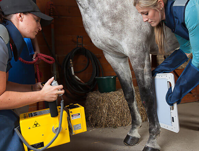
What are we talking about today? X-ray examinations of horses for sale. But first, a few completely casual, nameless, real-life cases that are 100% true.
🩺🐴 A 6-year-old horse undergoing a pre-purchase exam with hopeless sesamoid bones. Back when X-rays were graded by class, the vet assessed them as a strong Class III. For this reason, a female competitor decided against buying the horse and it ended up in different hands. The horse went on to participate in two Olympics, with its last Grand Prix competitions at the age of 19, comfortably completing an entire season of international competitions across several countries.
🩺🐴 A 5-year-old horse also with unattractive sesamoids full of nutrient canals, plus small and steep hooves, and showing a slight positive on flexion tests. Not looking optimistic… Yet in adulthood, this horse became something of a celebrity, winning international competition series one after another. At 17 years old, the mount was still vigorously completing M-level test with excellent results.
🩺🐴 A 3.5-year-old mare purchased as a sport prospect, examined from head to toe with a million X-rays, all crystal clear. After being transported to the new stable, the horse developed a not-too-large splint on the splint bone while in the paddock – „no big deal” really, but the periosteum wouldn’t calm down and week after week it continued to grow slightly. With conservative treatment failing, surgery was decided upon, with removal of the splint bone performed by the best surgeon at a renowned German clinic. Also „no big deal,” at least theoretically. Unfortunately, a complication after surgery was that the periosteum still wouldn’t settle down, and the splint continued to grow week after week on the stump of the splint bone… The game of calming the situation lasted nearly two years, most of which the horse spent on hand walks and later in a micro-paddock. In the end, to make it even more amusing, the horse not only returned to full sport (Medium/Advanced Level Dressage), but the periosteum flattened out, the splint was absorbed, and after another two years, there was no trace of the whole affair.
🩺🐴 A 7-year-old gelding in training. As the only horse of the rider (all her mounts were in active sport training from C-level upward), it passed a complete pre-purchase exam without objections, with good X-rays. The catch was that as a foal, the horse had a serious accident, falling into farm machinery that trapped him for many hours. He emerged from this unpleasant adventure with a broken splint bone, a fractured and displaced (or underdeveloped after fracture) end of one ischial tuberosity, and a fracture of the coffin bone. All of these healed spontaneously, and the horse started training and work as a 5-year-old. He was never lame; on flexion tests, on the lunge, and on various surfaces he was sound. Under saddle, he jumped, performed flying changes in series, and eagerly attempted piaffe. Nobody routinely X-rays splint bones, yet there was a complete fracture visible, with periosteal flooding and adhesion of the detached fragment. The missing half of the buttock (not supported by the ischial bone) was visible to the naked eye, but only if you stood directly behind the horse, facing the tail, comparing both buttocks—something nobody typically assesses during pre-purchase exams. The line from the old coffin bone fracture fell perfectly under the edge of the horseshoe; it was only visible on X-rays when the horse was barefoot. As long as the horse wore shoes, there was no chance of detecting this artifact. Either way, the horse moved well, wasn’t bothered by anything, and lacked nothing for riding at the M-level or small tour. However, if someone wanted to evaluate this mount solely by focusing on X-rays, I don’t think he would ever have received the green light for anything more than walking rides under saddle.
I could write down several dozen such stories right off the top of my head. No complete set of x-rays has ever provided any guarantee for a horse’s future performance.
Meanwhile, X-rays still remain crucial for many buyers and in many, if not most cases, they determine whether the decision to purchase a horse will be made.
How correct this approach is, is increasingly being verified by research. In England, a group of 300 adult and working riding horses was tested, each of which had some changes and deviations from the norm identified in X-rays. From this group, 92% of horses turned out to be clinically normal, without any lameness or other ailments. In the Netherlands, 1231 clinically healthy KWPN horses were examined, and in this group, almost half (44.3%) showed changes in joints resulting from osteochondrosis.
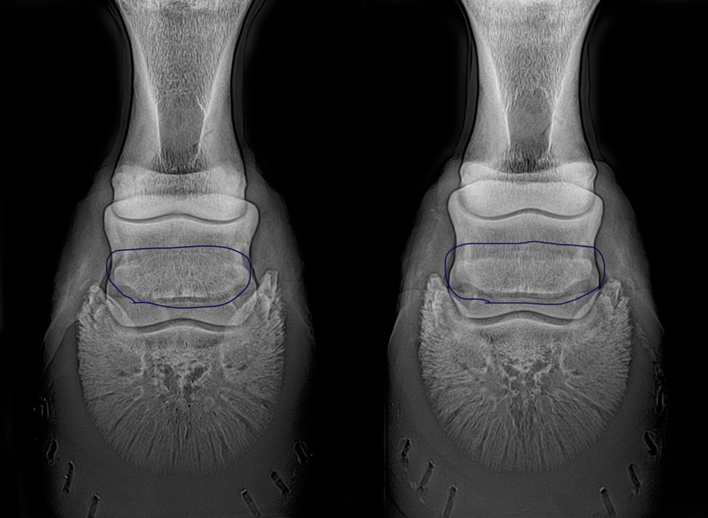
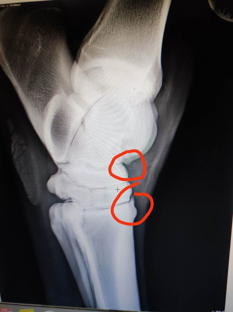
The usefulness of performing neck X-rays during pre-purchase examinations in non-hospital settings is currently being questioned, as portable devices don’t provide an image clear enough to make long-term assessments. Similarly with sesamoid bone images. Just a few years ago, changes in this bone would immediately be labeled as sesamoiditis. A rough edge, small hole, or channel, and suddenly the horse was considered ill. Currently, it’s believed that X-rays alone have no diagnostic value. And for clinically healthy horses, it’s impossible to determine from such images whether or when any health issues will develop.
Moreover, there’s actually little evidence that any finding on an X-ray will in any way affect the horse’s usability: „(…) deviations from the normal in neck and back X-rays are of no predictive value for future soundness or athletic potential,” as Horse and Hound magazine quoted from a Royal Veterinary College publication.
Of course, many changes observed in X-rays of equine limbs provide a very reliable basis for assessing the current condition and progression of the horse’s health, but „buyers should remember that there are still many deviations seen in limb X-rays that are not indicators of future problems.” Evaluating these changes requires extensive knowledge and practice, and hasty judgment can and unfortunately often does lead to unnecessarily rejecting a horse purchase.
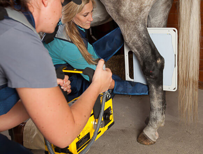
Year after year, examinations become increasingly meticulous, detailed, and comprehensive. Once, a standard 12 X-rays were taken; today it’s 40, and sometimes even 80. Equipment is becoming more technologically advanced, images more distinct, sharp, and contrasted (not to mention that they used to be produced on film, and today are captured with increasingly sensitive digital equipment). They will clearly show every shadow, every even slightest deviation from the norm. Something that until recently wouldn’t have even been a matter for discussion might now be considered a change in bone density and completely eliminate a horse from a potential sale/purchase.
What do the competitors say about this?
Firstly, every trainer I ask honestly admits that many of the horses they trained and sold 20 or even 10 years ago, which successfully competed in sports after being sold, wouldn’t pass examinations today. Not to mention that 20 years ago in Poland, nobody typically removed chips from horses’ joints. Sometimes a horse would have its first X-ray at age 10, revealing surprises that left everyone shocked – everyone except the horse, who was busy racing around and collecting trophies from competitions.
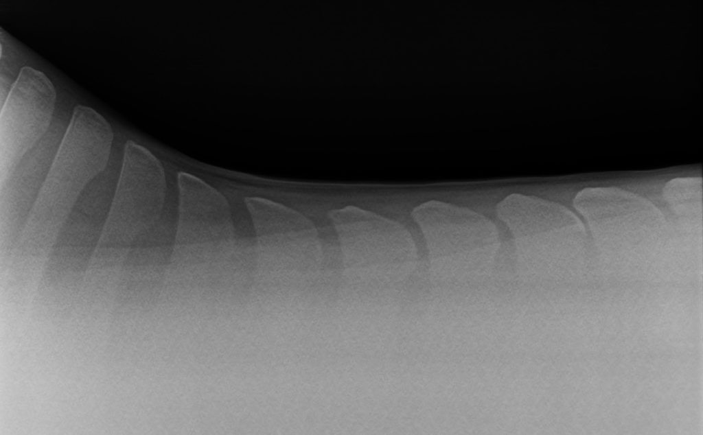
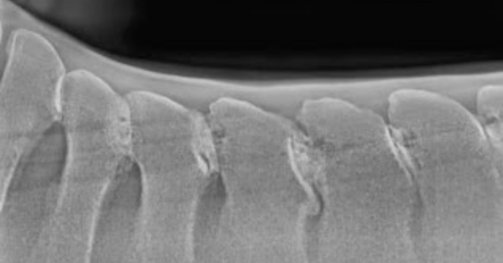
Secondly, it’s interesting that none of the professional athletes I know are restrictive about X-ray perfection. They don’t absolutely require the horse to be a walking anatomical atlas. Such demands, however, very often come from amateurs, people who buy „one horse in a lifetime” and can reject not just a few, but a dozen horses that have some changes and are therefore „unsuitable” for purchase and future use plans. X-rays of backs particularly lead this trend, due to what the industry calls self-diagnosis. Images of a horse’s thoracic spine (not cervical! nothing can be seen there anyway) are easy for amateurs to interpret. Whether the vertebrae touch each other or not, anyone can see and independently diagnose kissing spines.
Moreover, a veterinarian who says that such images with closely positioned vertebrae don’t necessarily have any significance will be perceived as incompetent. Because it’s visible to the naked eye! Meanwhile, many horses whose spinous processes overlap or touch show no clinical symptoms throughout their entire lives. But in pre-purchase examinations, to put it crudely – they get thrown in the trash.
What do veterinarians say about this?
Fortunately, more and more veterinary authorities are beginning to emphasize that X-rays cannot be evaluated „in a vacuum,” meaning without clinical examination and assessment of the horse itself. Unfortunately, veterinarians are often asked for such opinions, and it’s understandable that they prefer to express themselves cautiously, especially in the context of current legal regulations, existing warranties, and the genuine uncertainty about a horse’s future usability. It’s sometimes safer to „err on the side of caution” or reject a horse and avoid potential consequences in disputes and possible returns of the mount. As long as living animals are treated under the law like washing machines or refrigerators, which are expected to maintain their precisely defined properties for years, this will continue to favor more restrictive radiological assessments.
What’s also specific is the fact that potential buyers, often lacking detailed knowledge or experience with horse use, panic at the news of a change or deviation from the norm on an X-ray and don’t allow much room for explanation. Because if „something is wrong,” they assume the entire horse must be a ticking time bomb that will become lame at any moment. Here, the veterinarian’s communication skills matter more than the clinical report itself. If they can clearly and vividly explain what’s actually on the X-rays, what might be significant, and what definitely has no importance, there’s a chance the potential buyer won’t run away screaming.
Perhaps the best description I’ve encountered regarding a completely insignificant yet visible change on an X-ray was comparing it to a crack in the plaster at the junction of two drywall sheets. Yes, you can see such a crack and can’t pretend it’s not there, but it’s not a structural defect and certainly won’t cause the house to collapse. It won’t make the wall leak or the room get cold. In reality, the biggest problem with it is that… it looks unattractive.
What do potential buyers say about this?
Potential buyers want perfect X-rays. Some buy horses at auctions or from abroad, remotely, based on videos – in such cases, a complete set of X-rays is essentially the first stage of selecting potentially interesting horses. Others keep in mind the possibility of reselling the horse in the future (and since buyers want perfect X-rays…). Still others assume from the start that any deviation from perfection means that there is „something wrong” with the horse, and they don’t want to deal with such a defective mount on principle. Of course, having requirements is every client’s right, but the fact that some record-holders manage to X-ray 15-20 horses and reject each one as absolutely unacceptable should give us pause for thought…
Conclusions
I definitely advocate giving a chance to horses that aren’t perfect and absolutely not expecting perfection in complete sets of X-rays. Perfection is not the norm in X-rays – minor deviations, small asymmetries, slight imperfections – that is the actual norm.
Indeed, X-rays can be invaluable and decisive in many cases – for example, when diagnosing an existing lameness in a horse, or when we know it has some deficiency and want to check and monitor it over time to see if the condition is growing or remaining unchanged. Sometimes such documentation can even defend an „ugly X-ray” if the horse works, competes, and its X-ray remains just as ugly 😅 for several years.
If we expect a horse with perfectly flawless X-rays across the entire set, we may never find one.
At some point, we definitely need to ask ourselves whether we want to buy good horses or good X-rays. Personally, I prefer horses 😉
Dodaj komentarz